Research themes
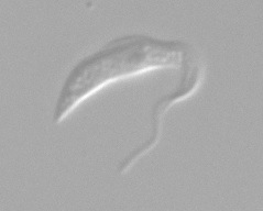
A T. brucei cell, visualised by differential interference contrast microscopy
1. The cytoskeleton of Trypanosoma brucei
The cytoskeleton of trypanosomes is defined by a corset of subpellicular microtubules. We investigate how the dynamics of these microtubule arrays are regulated and contribute to morphological adaptations during the life cycle of the parasite. A particular focus is on the role of microtubule posttranslational modifications, such as polyglutamylation. This is a joint project with the Department of Experimental Physics (Prof. Matthias Weiss), funded by the DFG priority program "Physics of Parasitism".
We use a variety of molecular biology techniques, such as mutagenesis, epitope-tagging, gene deletions and RNA interference. Equally important are, however, cell biology techniques to probe the 3-dimensional structure of the cells. Techniques such as immunofluorescence microscopy, advanced digital image analysis (cell tracking, motility analysis) and electron microscopy are routinely employed.
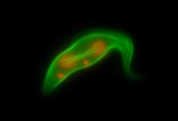
The mitotic spindle stained with anti-tubulin antibody KMX (see Ogbadoyi et al., Chromosoma, 2000)
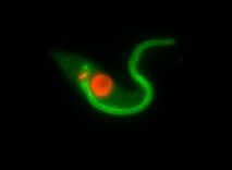
A calpain-like protein localises to the flagellum of T. brucei (see Liu et al. MBP, 2010).
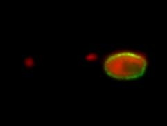
NUP1, a protein of the nuclear envelope in T. brucei (see Ogbadoyi et al., Chromosoma, 2000)